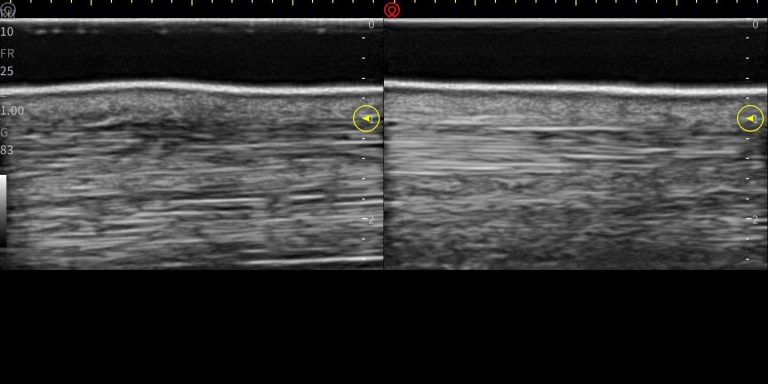
What is B Mode ultrasound
Basic Principle of Ultrasound Imaging
Ultrasound is identical that of Sonar technology used at sea, where Sound wave produced using Piezo electric crystal having property, for a given electrical pulse to produce sound wave with range of frequency. This Depends on its crystalline chemical property, size & thickness as well as the overall layout of the electrically sensitive ceramic crystals. Design and numbers of crystals used depends on the required, examination type, size, distance, and property of the target material under examination. When crystal energised with short pulse of alternative current, individual crystals sends out pulse of wave containing sound energy by means of resonating frequency of vibration.
Hence sending out low or high frequency sound beam or a pressure wave in a definite direction.
This is through a coupling medium, or for medical examinations Ultrasound gel used as a coupling for waves to transfer into the body with minimum attenuation.
The reflected and returning resultant wave is detected by the same crystal, having property of mechanical pressure to be converted into electrical pulse, energy contained in this wave is in sympathy of the returning sound waves containing information of reflective layer’s distance in terms of time, attenuation, refraction and deflection of intermediate layers and reflectivity in terms of reduced amplitude of the incidental sound wave. Detected wave amplified and processed further. makes a single line of echo. Historically known as A-Gate or A-mode Ultrasound
Basics of B mode Ultrasound
Depending on type of examination, access or contact required, type of crystal, Shape and number of crystals used to produce individual echo lines. With synchronised timing and processing of individual ultrasound echo lines put together
B Mode Ultrasound Image created by transmission of sound waves in the body. Geathering overall information from reflected echo lines in terms of instantaneous amplitude, returning frequencies in relation to depth related returning time from body. Shape of cross-sectional ultrasound image depends on all crystals arranged in Curved or Linear shapes.

Preprocessing
Preprocessing used to synchronise transmitting and receiving lines from all the crystals, for a required depth to multiplex and energise the crystal to achieve focused cross-sectional slice within the returning echo, carrying information of the field under examination. Having exact delay time, like radar systems each dot to display as deferent grey value to other. This is to complete a raw image that depends on the Intensity of the returning echo signals.
Post processing
Image optimisation controlled to improve visual quality and for interpretational diagnostics quality by the user depending on the user requirements is post processing. Various controls enable user to optimise returning echo like increasing gain for amplification of returning echo resulting in higher contrast or reduction to reduce saturation. TGC (time gain compensation) to control echo at various depth in terms of time delay gain control. Frequency of transmission or returning echo. Focus by getting more information at given depth, choosing dynamic range of greys by increasing or decreasing grey value divisions. Averaging in formation from adjourning echo lines, or of various algorithms values from previous cross sectional frames frame filtering or by higher or lower persistence keeping constant information more and updating new only.
Quality of the image:
Image information depends on
1 selecting correct scanner, and transducer
Due to its design &, property of the scanner, transducer's shape, crystal, size, spacing and numbers. To produce finer information from physically possible maximum number of echo lines Also depends on the probe or transducer’s physical dimensions and the requirement of an examination to be performed.
All above effects processed by scanner's receiver for transmission and amplification of echo, filtering analogue signal in terms of amplification, frequency filtering and noise, followed by digitisation and Scan conversion in relation to time synchronisation. These depends on quality of the Ultrasound technology electronic design. Choice of these, with pre and post processin and ultimatly user's skill for optimisation and anatomical knowldge provides very versatile and economical method for soft tissue anatomical examination and diagnosis.
At demonstrations or at delivery and installation time, detailed training is always provided to our customers.
Applications & User Presets:
Overall Ultrasound configurations and Sets of User selected Parameters with types of examinations, selection of gain, frequency depth, focus, dynamic range, persistence, grey map, filtering, angle width, orientation, size, power, are called User Presets. These provides easy and fast methods for individual probes and related variations of possible applications settings. Once added with clinical side of anatomical, measurements, & calculations requirements in individual repeatable choices of sets User can save time and optimise image just to optimise for any variations between patients or in terms of size or location of the anatomy under examination
We need your consent to load the translations
We use a third-party service to translate the website content that may collect data about your activity. Please review the details in the privacy policy and accept the service to view the translations.

