

In the field of Small Animal veterinary, none invasive abdominal examination by Ultrasound imaging plays a crucial role in diagnosing various conditions. Pet's health coditions related to the liver, bladder, and kidneys in cats. by low cost, non-invasive ultrasound imaging technique allows veterinarians to obtain detailed images of these organs, aiding in the detection of abnormalities and guiding appropriate treatment plans.
When it comes to the liver, abdominal imaging by ultrasound scanners helps identify liver diseases such as hepatitis, cirrhosis, and tumors. By visualizing the liver's size, shape, and texture, veterinarians can assess its overall health and function. Similarly, US imaging of the bladder allows for the detection of urinary bladder stones, tumors, or infections that may be causing urinary issues in cats.
Furthermore, cat's kidney by ultrasound imaging is essential in evaluating renal health and detecting conditions like kidney stones, cysts, or tumors. By examining the size, shape, and structure of the kidneys, veterinarians can assess their function and identify any abnormalities that may require further investigation or treatment.

Canine ultrasound imaging is a valuable diagnostic tool used in small animal veterinary scanners to examine various organs and conditions in dogs.
When it comes to abdominal ultrasound, the scanner allows veterinarians to obtain detailed images of the dog's liver, kidney, bladder, pancreas, spleen, and other organs. This non-invasive technique helps identify abnormalities such as masses, cysts, or inflammation, aiding in the diagnosis and treatment of potential health issues.
In addition to abdominal imaging, ultrasound is also used to assess the heart and cardiac function in canines. This is particularly important for larger breeds that may be prone to certain heart conditions. By visualizing the heart's structure and blood flow, veterinarians can detect abnormalities like valve defects, cardiomyopathy, or fluid accumulation, enabling appropriate intervention.
Overall, canine ultrasound imaging plays a crucial role in veterinary medicine, allowing for early detection and monitoring of various conditions. It helps veterinarians provide more accurate diagnoses and develop effective treatment plans for our beloved furry companions.

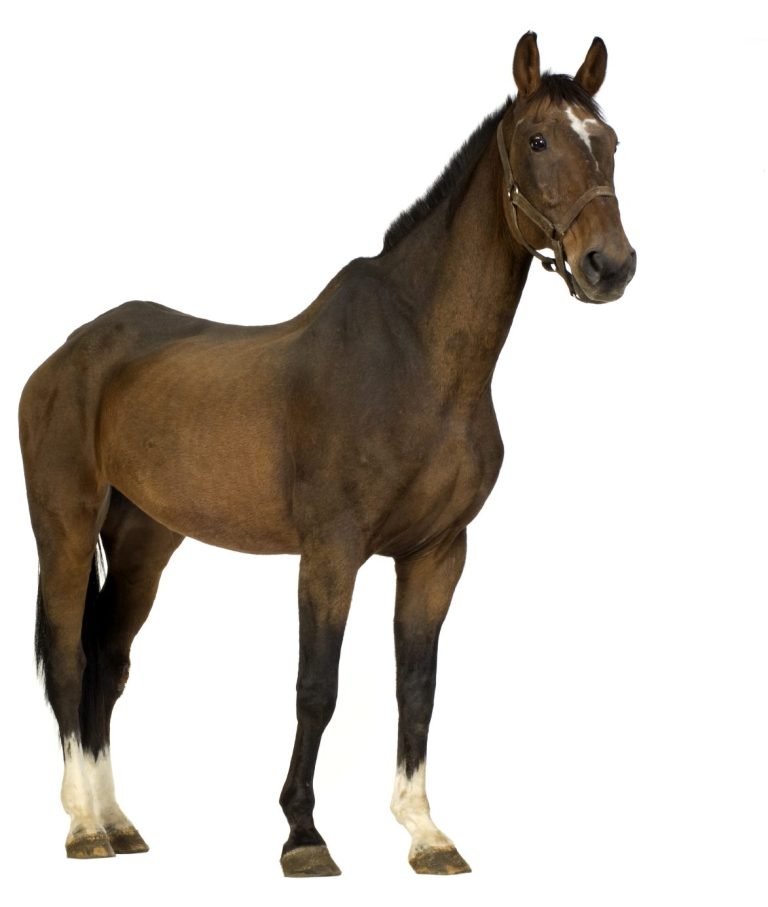
Equine reproduction is a fascinating field that plays a crucial role in the breeding and management of horses. Portable ultrasound scanners have revolutionized the way we approach equine reproductive examinations.
One of the key advantages of portable ultrasound scanners is their lightweight design. This makes them highly convenient and easy to transport, allowing veterinarians to perform reproductive exams in various locations. Whether it's at the stud farm for equine pregnancy, fertility scans for mares or for tendon exams prior or during examinations for competitions, Pir Data Ultrasound scanners provide flexibility and accessibility.
Furthermore, portable ultrasound scanners from Pie Data UK offer exceptional image quality. This is essential when evaluating the reproductive system of mares. With high-resolution imaging, veterinarians can accurately assess the ovaries, uterus, and other reproductive structures. This enables the detection of abnormalities, such as cysts or tumors, which may impact fertility. Examining Ovarian stages of initial fertilisation and corpus luteum ultrasound is a common procedure in equine reproduction for early pregnancy detection. By using portable scanners, veterinarians can easily visualize and measure these structures. This information is vital in determining the mare's breeding suitability and optimizing reproductive health management.
Portable ultrasound scanners also play a crucial role in diagnosing reproductive pathological normality and monitoring pregnancy. They allow for early detection of conditions like endometriosis or uterine infections, enabling timely treatment. Additionally, these scanners provide real-time monitoring of fetal development, ensuring the health and well-being of both mare and foal.
Equine tendon injuries pose a significant concern within the owners. These injuries commonly arise from the strains associated with sports or excessive exertion in horses. In cases where such an injury is suspected, a thorough expert veterinary investigation becomes imperative to accurately diagnose the extent of the ligament damage. One of the most readily available tool to veterinarian is portable ultrasound scanner.
The duration of healing for equine tendon injuries varies contingent upon the severity of the harm inflicted. Consequently, it becomes paramount to closely monitor the progress of the healing process to ensure optimal recovery. Regular follow-up examinations utilizing veterinary ultrasound scanners are indispensable for assessing advancements and making any necessary modifications to the treatment regimen. Exercise assumes a pivotal role in rehabilitating equine tendon injuries.
However, striking an equilibrium between rest and activity is crucial to prevent exacerbating the injury. Veterinary scanners prove invaluable in tracking the healing process and determining the appropriate level of exercise required for the horse's recovery.
+441293510231
©Copyright Pie Data UK Ltd 2023. All rights reserved.

We need your consent to load the translations
We use a third-party service to translate the website content that may collect data about your activity. Please review the details in the privacy policy and accept the service to view the translations.